Salinity Stress Alters Nutrient Uptake and Causes the Damage of Root and Leaf Anatomy in Maize
DOI:
https://doi.org/10.18502/kls.v3i4.708Abstract
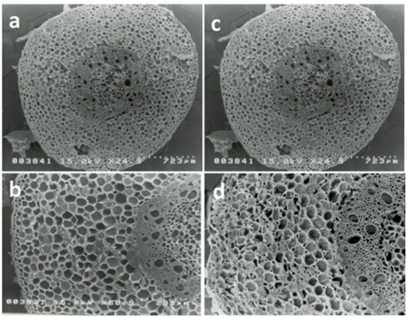
Salinity is one of major problems in agriculture especially in arid and semiarid area due to causes the damage of many aspects in plant growth and development. This study observed root and leaf anatomy and nutrient uptake in maize plants exposed to salinity stress. Maize seedlings were placed in the plantation room under same temperature, humidity and light intensity conditions and were treated with 0 %, 1 %, 2 % and 3 % NaCl for 5 d. Anatomy of root and leaf were observed using scanning electron microscopy (SEM). Nutrient uptake was estimated by the content of trace elements of leaves. Trace element were quantified using inductively coupled plasma-mass spectrometry (ICP-MS), but chlorine was determined by an atomic absorption flame spectrometer. The results showed that salinity slightly damaged roots anatomy. Epidermis cells and parenchyma cells of cortex and pith were shrinkage in 2 % and 3 % NaCl-treated plants. Leaf anatomy showed mesophyll and bundle sheath cells which slightly suppressed. Meanwhile, chloroplasts content inside those cells were dramatically decreased. Anatomical damage of roots and leaves was accompanied by altering uptake of some trace elements. The contents of aluminum, calcium, iron, magnesium, sodium, chlorine, in NaCl-treated plants were higher than control. Otherwise, boron, potassium and phosphor were lower in NaCl-reated plants. The rest of trace elements were in comparable concentration.
Keywords: maize; leaf; nutrient; root; salinity
References
FAO., Global network on integrated soil management for sustainable use of saltaffected soils. Land and plant nutrition management service; 2000. Accessed in 5 April 2014. Available on: http://www.fao.org/ag/agl/agll/spush.
R. Munns, “Genes and salt tolerance: Bringing them together,” New Phytologist, vol. 167, no. 3, pp. 645–663, 2005.
R. Munns, “Comparative physiology of salt and water stress,” Plant, Cell and Environment, vol. 25, no. 2, pp. 239–250, 2002.
M. A. Hajibagheri and T. J. Flowers, “X-ray microanalysis of ion distribution within root cortical cells of the halophyte Suaeda maritima (L.) Dum.,” Planta, vol. 177, no. 1, pp. 131–134, 1989.
T. J. Flowers and M. A. Hajibagheri, “Salinity tolerance in Hordeum vulgare: Ion concentrations in root cells of cultivars differing in salt tolerance,” Plant and Soil, vol. 231, no. 1, pp. 1–9, 2001.
P. M. Hasegawa, R. A. Bressan, J.-K. Zhu, and H. J. Bohnert, “Plant cellular and molecular responses to high salinity,” Annual Review of Plant Biology, vol. 51, pp. 463–499, 2000.
T. J. Flowers and A. R. Yeo, “Ion relations of plants under drought and salinity.,” Australian Journal of Plant Physiology, vol. 13, no. 1, pp. 75–91, 1986.
H. Greenway and R. Munns, “Mechanisms of Salt Tolerance in Nonhalophytes,” Annual Review of Plant Physiology, vol. 31, no. 1, pp. 149–190, 1980.
R. Munns and A. Termaat, “Whole-plant responses to salinity.,” Australian Journal of Plant Physiology, vol. 13, no. 1, pp. 143–160, 1986.
X. Niu, R. A. Bressan, P. M. Hasegawa, and J. M. Pardo, “Ion homeostasis in NaCI stress environments,” Plant Physiology, vol. 109, no. 3, pp. 735–742, 1995.
J. A. Hernández, E. Olmos, F. J. Corpas, F. Sevilla, and L. A. del Río, “Salt-induced oxidative stress in chloroplasts of pea plants,” Plant Science, vol. 105, no. 2, pp. 151– 167, 1995.
D. A. Meloni, M. A. Oliva, C. A. Martinez, and J. Cambraia, “Photosynthesis and activity of superoxide dismutase, peroxidase and glutathione reductase in cotton under salt stress,” Environmental and Experimental Botany, vol. 49, no. 1, pp. 69–76, 2003.
R. K. Sairam and G. C. Srivastava, “Changes in antioxidant activity in sub-cellular fractions of tolerant and susceptible wheat genotypes in response to long term salt stress,” Plant Science, vol. 162, no. 6, pp. 897–904, 2002.
J. M. Gómez, A. Jiménez, E. Olmos, and F. Sevilla, “Location and effects of long-term NaCl stress on superoxide dismutase and ascorbate peroxidase isoenzymes of pea (Pisum sativum cv. Puget) chloroplasts,” Journal of Experimental Botany, vol. 55, no. 394, pp. 119–130, 2004.
S. Rahman, T. Matsumuro, H. Miyake, and Y. Takeoka, “Salinity-induced ultrastructural alterations in leaf cells of rice (Oryza sativa L.),” Plant Production Science, vol. 3, no. 4, pp. 422–429, 2000.
S. Mitsuya, M. Kawasaki, M. Taniguchi, and H. Miyake, “Light dependency of salinity-induced chloroplast degradation,” Plant Production Science, vol. 6, no. 3, pp. 219–223, 2003.
K. Yamane, M. Kawasaki, M. Taniguchi, and H. Miyake, “Differential effect of NaCl and polyethylene glycol on the ultrastructure of chloroplasts in rice seedlings,” Journal of Plant Physiology, vol. 160, no. 5, pp. 573–575, 2003.
R. Hasan, Y. Ohnuki, M. Kawasaki, M. Taniguchi, and H. Miyake, “Differential sensitivity of chloroplasts in mesophyll and bundle sheath cells in maize, an NADPmalic enzyme-type C4 plant, to salinity stress,” Plant Production Science, vol. 8, no. 5, pp. 567–577, 2005.
R. Hasan, M. Kawasaki, M. Taniguchi, and H. Miyake, “Salinity stress induces granal development in bundle sheath chloroplasts of maize, an NADP-malic enzyme-type C4 plant,” Plant Production Science, vol. 9, no. 3, pp. 256–265, 2006.
K. M. Volkmar, Y. Hu, and H. Steppuhn, “Physiological responses of plants to salinity: A review,” Canadian Journal of Plant Science, vol. 78, no. 1, pp. 19–27, 1998.
T. J. Flowers and A. R. Yeo, “Ion relations of plant under drought and salinity,” Aust. J. Plant Physiol., vol. 13, pp. 75–91, 1986.